• Suite #3. Pine Medical Centre, 3rd Avenue Belleville. St Michael, Barbados
(246) 427 5863

Category Archives: Uncategorized

GRAVITATIONAL ECZEMA

This is an inflammatory condition of one or both of the lower extremities due to insufficiency of the leg veins. It is also known as stasis dermatitis and venous eczema.


Valves in the deep, perforator and superficial veins help blood to flow from the lower limbs to the heart efficiently. Incompetent valves allow flow in the opposite direction and this increases venous pressure, which results in fluid leaking into the surrounding tissues. This then triggers inflammation in the skin.


Gravitational eczema is more common in middle-aged patients with females being more commonly affected. Smoking, obesity, pregnancy and inactivity associated with excessive sitting or standing are aggravating factors. Acquired venous insufficiency due to surgery, trauma and deep vein thrombosis can precipitate this condition.


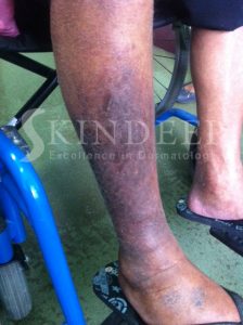
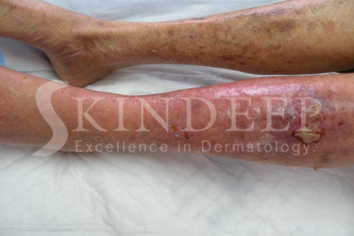
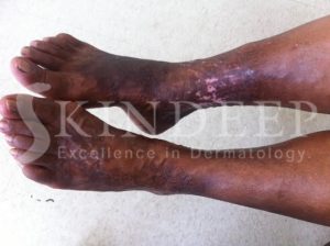
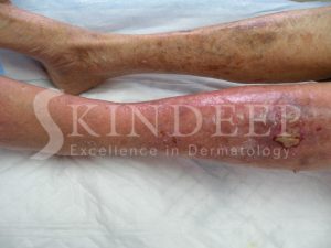
The earliest symptom is increasing swelling of the ankles during the day, which  resolves with lying in bed while sleeping. This becomes more severe and constant and is associated with aching discomfort and varicose veins. A dark pigmentation then appears just above the ankle due to haemosiderin deposition in the tissues from the breakdown of red blood cells. Eczema changes range from itchy red papules and blisters (acute) to dark thickened, hard, cracked and peeling skin (chronic) either in patches or generally on the lower two thirds of the leg. Net like or poly angular atrophic white scars with red bumps are sometimes present, especially on the lower inner aspect of the ankle, and these are called atrophie blanche.


Lipodermatosclerosis is the name given to the chronic changes associated with chronic venous insufficiency. These changes consist of haemosiderin pigmentation, atrophie blanche and a hardening/thickening of the lower third of the legs due to inflammation of the subcutaneous fat. The leg takes on an “inverted champagne bottle” or rolling pin appearance that is permanent.


Complications of venous eczema include infection, which can take the form of a weeping honey coloured liquid associated with blisters and dark crusts called impetigo. Spreading superficial skin infection associated with warmth, redness and swelling is called cellulitis. Contact dermatitis can also occur due to the various medications and dressings used during treatment. 


The most feared complication however is the venous ulcer that can be chronic and cause significant emotional distress as well as decreased quality of life.


It is important to keep physically active and to avoid standing for long periods without engaging the calf muscles as a pump to return blood to the heart. While sitting or lying down the legs should be elevated above the hips. The use of gentle soaps and moisturisers allows for proper surveillance and detection of complications. Compression bandages and hosiery are required to limit the swelling from vein incompetence. Above all, trauma must be avoided as it can precipitate ulceration.


Oral medications are used to improve vein circulation like pentoxifylline. Patients with a history of deep vein thrombosis are kept on oral prophylactic medication and generally infection is treated with topical or oral antibiotics. Wet eczemas and infections require wet compresses, with potassium permanganate or diluted white vinegar, and respond to steroid creams. Chronic eczematous changes respond better to greasy moisturisers and steroid ointments.  


Ulcers are kept clean and dressed and in some cases physical methods like vacuum dressings or hyperbaric oxygen are used. Surgical methods are very effective including skin grafts, flap closure, laser ablation, endoscopic vein surgery and sclerotherapy to name a few.


In our country where obesity and diabetes is so common, stasis dermatitis and subsequent ulcer formation can contribute to loss of limb, disability and morbidity. Since it is largely preventable, we must all do our part via lifestyle changes including diet and exercise to reduce the burden of care on our health budget.


Early intervention by your doctor is critical to enable positive outcomes.

FRECKLES

Freckles are small 1-3 mm pigmented spots which appear predominantly on the face and other sun exposed areas.


They represent pigment produced by the skin pigment cells called melanocytes which then feed it to the surface cells called keratinocytes.

The ability to form freckles depends on your genetics and the type of skin given to you by your parents. lighter skin types which burn more easily manifest freckles as a sign of skin damage caused by the sun. Some individuals with darker skin can also manifest freckles. Sunburns and the tendency to use insufficient sunblock or working outdoors can predispose to this condition.


They can be present in the winter and fade or disappear in the summer.


It is important to realize that freckles represent damage of the skin from the sun.

FERRITIN AND YOUR HAIR

The average human scalp has 100,000 to 150,000 hairs and loses 80 to 100 per day. The hair cycle consists of three parts. Anagen lasts for 3 to 6 years and is the phase of hair growth that determines the length of your hair.  Catagen is a transitional stage between hair growth and hair fall, which lasts about two to three weeks. Telogen refers to the stage of hair fall and inactivity of the follicles that lasts about three months. 


Diffuse hair loss can occur if hair growth or the length of anagen is decreased or the normal telogen percentage is increased. This results in extra shedding of hair in the shower, on clothes, combs, brushes and pillows. A balanced diet containing protein, iron and nutrients (like zinc, magnesium, Vitamin A & D, biotin and selenium) is required for healthy hair growth.


One of the most common causes of hair loss in pre-menopausal women is reduced iron storage in the body measured by the plasma ferritin. Iron is an important constituent of Haemoglobin and myoglobin, which are the oxygen carrying molecules in the blood and muscles respectively. Ferritin stores iron and releases it in a controlled manner. It is stored in the cells of the body and found in the blood but the main storage areas are the liver, spleen, skeletal muscles and bone marrow. 


An imbalance in the absorption of iron compared to the quantity lost via excretion can lead to an excess or deficiency. In the latter case your iron stores or ferritin is utilised to make up the shortfall. Initially your haemoglobin will remain normal and no signs of anaemia will be evident but as ferritin levels fall further, the storage levels become insufficient to maintain normal haemoglobin levels and anaemia ensues. Long before that happens diffuse hair loss occurs at ferritin levels below 70ng/ml and gradually gets worse. Normal serum ferritin levels range from 24-336ng/ml for men and 11-307ng /ml for women. These normal limits vary from one lab to another.


Besides being a possible trigger for telogen effluvium where a lot of hair shedding occurs, iron deficiency could also be the most likely underlying cause when hair loss is subtle; slowly thinning out over many months or even years. It is therefore evident that iron is essential for the normal growth and maintenance of hair. A normal haemoglobin level does not mean that your iron stores are sufficient to support healthy hair growth.


Menstruation and pregnancy make premenopausal women most vulnerable to iron deficiency. Poor diet, chronic illness, poor digestion and loss via gastrointestinal bleeding can also be contributory factors. Tannins found in tea and coffee, bran and egg albumin can inhibit iron absorption and contribute to deficiency.


The good news is that hair loss due to low ferritin is entirely reversible, and relatively easy to correct. After the ferritin level has been ascertained by means of a blood test, attention to your diet is the first step to recovery. Eating a combination of iron-rich foods such as red meat, green, leafy vegetables, shellfish, egg yolk, nuts and cereals with food or drinks that are high in vitamin C For example, a glass of freshly squeezed orange juice will maximize your iron absorption. Iron supplements in the form of ferrous fumarate or ferrous sulfate can be prescribed to speed up the process or in severe cases.


No matter what your cause of hair loss, it is important to realize that low ferritin levels can aggravate your problem. See your Dermatologist to determine the exact cause of hair loss.

ECCRINE HIDROCYSTOMA

Sweat glands are widely distributed on the body and each gland consists of a single duct and a coiled deeper component. These benign tumours of the eccrine sweat gland form cystic swellings of the sweat duct adjacent to and around the eyelids. They may be multiple (Robinson type) or solitary (Smith and Chernosky type). Another sweat gland forming cystic swellings associated with the eyelids are apocrine glands but they occur on the lower eyelid margin and eyelashes. 


Eccrine hidrocystomas are prevalent between the ages of 30 and 70 years of age. Even though they are said to be rare, local dermatologists see one or two cases every year. Generally both males and females are affected but it is more commonly seen in females. 


Dome shaped growths ranging from 1-6 mm are observed. In black patients they are skin coloured but in Caucasians they may appear amber, brown or bluish. In hot conditions like ours, these lesions get larger and can seem to multiply. This is due to the accumulation of sweat, which causes the structures to dilate. In cool conditions they become smaller so that really tiny ones almost disappear. Eccrine lesions do not involve the eyelid margin but are distributed around the eyelid skin unilaterally or bilaterally.

The desire for cosmetic improvement often drives the desire for treatment, as the condition is not dangerous. Temporary relief can be achieved by simple needle puncture under sterile conditions. Needle puncture is only practical in cases of small solitary hidrocystomas and recurrence is almost inevitable.


The treatments of multiple lesions using 1% atropine or scopolamine creams, has been successful. Unfortunately the anticholinergic side effects such as dryness of the mouth, eye irritation or flushing of the face can occur.

Electrosurgery of the base of the lesion after careful removal of the exterior lining is the method of choice in Barbados. The occurrence of scars is uncommon and the recurrence rate is low. Surgical excision followed by stitching is not recommended due to the formation of scars. Carbon dioxide and pulse dye lasers show promise and may be the treatments of the future.


Due to the diagnostic ambiguity there are some cases where a biopsy is required to rule out basal cell carcinoma, epidermal cysts, hemangiomas, lymphangiomas and mucoid cysts.

It is important to see your dermatologist or eye doctor to determine the nature of growths around the eyes and to determine the necessity for treatment. In the case of the Eccrine Hidrocystoma it is best to avoid hot temperatures or humid conditions.

DERMOGRAPHISM

Dermographism is a form of skin writing, which occurs in approximately 2-5% of the population. This is the most common form of physical urticaria and occurs when the skin is firmly stroked or rubbed. Capillary dilation produces an initial red line that is followed by arteriolar dilation resulting in broadening of the area. The final phase in this Triple Response of Lewis is the collection of fluid to form a wheal (Bajans call it a “whale”).


The exact cause and mechanism of dermographism is not known. Stroking of the skin results in the stimulation of mast cells and the subsequent release of histamine along with other inflammatory mediators in the affected area. Histamine causes the dilation of blood vessels making them leak fluids and also causes itching. Secondary causes include allergies to external agents like Penicillin, Scabies or worm infestation, insect bites and emotional upset. Patients with excessive mast cells (mastocytosis) can initially display skin writing tendencies early on as the disease progresses.


This condition can occur at any age but is more common in teenagers and young adults and there is no race or gender preference observed. Incidental trauma or pressure from chairs, rough collars/cuffs/seams, contact sports and towelling can be triggers. Hot environmental conditions, dry skin and any itchy dermatological condition e.g. eczema, can increase your risk. 


Within 5-10 minutes of a stroking stimulus a tingling, itchy red swelling occurs which represents the area previously stroked. These individual raised wheals/hives usually last for about 30 minutes. Itching and whealing can affect the entire body but the scalp and genitals are less frequently involved. The sensation of perpetual and chronic itching can be very disconcerting and be a cause of severe emotional distress. The ability to create elaborate patterns on the skin as a form of body art can be intriguing to observers and even appear supernatural! When objects are used to write words on the skin they magically appear a few minutes later.


Your Dermatologist can easily make this diagnosis from a thorough history and even though it may not be present at your visit, a simple test can elicit the signs. This test involves using an orange stick or blunt object to elicit dermographism. 


It is important to understand that this condition is not dangerous but more of a nuisance. Avoidance of trigger factors is imperative to reduce severity. Patients can be affected for a few weeks, months or years either continuously or at intervals. Antihistamines are very effective agents in the treatment of Dermographism. Phototherapy may be used in resistant cases.


The good news is it often resolves on its own.