• Suite #3. Pine Medical Centre, 3rd Avenue Belleville. St Michael, Barbados
(246) 427 5863

Author Archives: admin

VIRAL WARTS

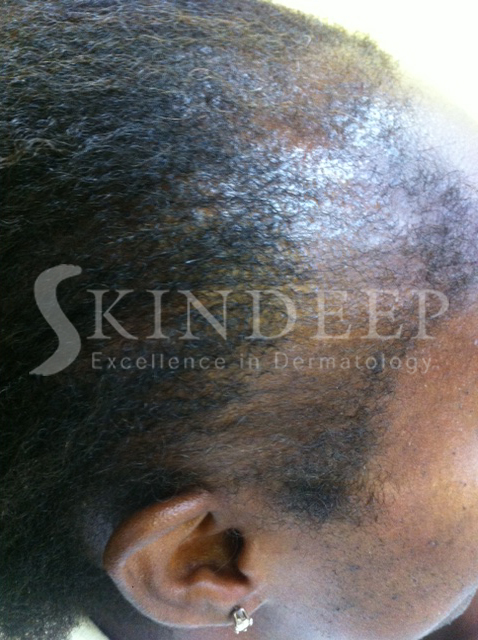
Viral warts are caused by infections with the Human Papillomavirus. There are at least 70 subtypes of this virus. Warts can appear anywhere on the body and commonly affect children and young adults even though all age groups are susceptible. Warts do not have “roots” and they only grow in the upper layer of the skin called the epidermis.


This infection is spread by skin-to-skin contact between individuals, autoinoculation, or from objects and surfaces. Skin that is damaged or wet and macerated is more susceptible to infection. Biting and sucking your fingers can also predispose you to warts. 


Communal bathing and changing facilities such as hotel rooms, bathrooms and swimming pools are areas where warts are contracted. Individuals with weakened immune systems or on some types of medication are also prone to chronic, stubborn episodes.


TYPES OF WARTS


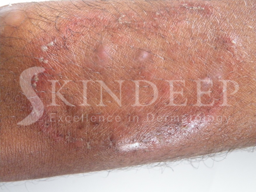
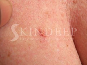
COMMON WARTS take the form of roughened skin coloured papules, which may be single or multiple. They are found most commonly on the dorsum of hands, toes, knees and elbows.


PLANTAR WARTS present as roughened flat areas, which penetrate deeply due to the pressure of your body weight and occur on the soles of the feet.

The MOSAIC pattern refers to clusters of warts over an area, which can be several centimetres wide. They can be painful and create difficulty with walking and are resistant to treatment.


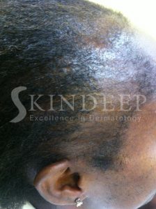
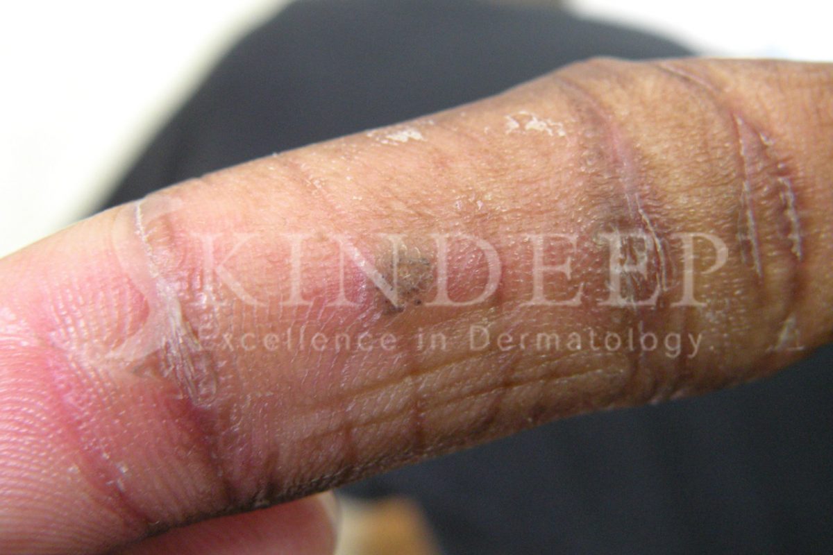
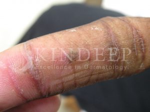
PLANE WARTS are usually 1-3 mm flat-topped papules that are smooth or have a slightly dry surface. The colour can vary from skin coloured to darker or lighter. The face and limbs are most often affected and they can be numerous. Shaving, incidental trauma and topical steroids spread them.


PERIUNGIAL WARTS occur around and under nails sometimes distorting nail growth. These warts can be stubborn as well.


FILIFORM WARTS usually present as finger like growths on a stalk.

GENITAL WARTS appear in various clinical forms that are transmitted sexually and can predispose to cervical, vulval and penile cancer. 


DIAGNOSIS


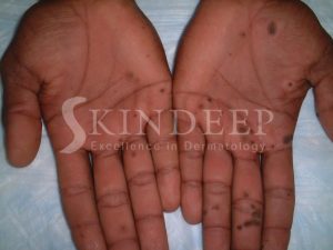
Warts are easy to diagnose clinically but there are some situations where corns, seborrhoeic keratoses or punctate keratodermas can cause confusion. Very often black dots are observed within a wart, which represent thrombosed blood vessels. Scraping or paring of a wart results in bleeding points, corresponding to these blood vessels. Scraping of a corn however, results in skin which looks progressively more normal. Corns are also painful to direct pressure.


TREATMENT


Left alone about 3 in ten warts disappear in ten weeks and even more resolve in 1-2 years.


There is a study that suggests covering common warts with adhesive/duct tape for six days a week, with the seventh used for soaking and paring down the exposed wart actually works. Results vary from encouraging to useless.


Salicylic acid wart paints can be used to treat warts in all areas except the face and genitals. It is best to soften the wart by soaking in warm soapy water, followed by paring with a pumice stone or emery board. The paint is applied accurately to the wart and then covered with plaster or duct tape. This process is repeated for approximately four months or until resolution.


Liquid nitrogen is applied to warts with a cotton bud or sprayed on to cause three or four freeze thawing cycles in one treatment. These treatments are performed every three to four weeks until the warts disappear. On average approximately one to four treatments are required but some stubborn warts require more. Aggressive freezing sessions can result in a blister that can be popped with a clean needle and treated with topical antibiotic. Over the counter spray freezing methods are less effective than liquid nitrogen. Combining freezing and cryotherapy is usually the best treatment.


In cases where a wart is stubborn and solitary (or just a few in number) a surgical approach can be used as a last resort. Unlike the other methods this results in a scar. The method most often used is curettage and electrosurgery in which the wart is scraped and scooped out with a curette before the wound is cauterised electrically. These wounds heal in about two weeks.


Lasers, cytotoxic drugs and immune modulators are also used to treat warts.


The spread of warts can be minimised by avoiding the sharing of towels, shoes as well as socks. When swimming, a waterproof plaster should be used to cover your wart. In the case of individuals with plantar warts, it is important to wear slippers in communal bathrooms and showers. Younger children should be discouraged from sucking fingers and biting nails if they already have warts.

TRACTION ALOPECIA

Traction alopecia is a form of non-scarring hair loss predominantly affecting the hairline and is due to prolonged tension on the hair follicles. The condition was first described in 1907, by the Austrian dermatologist Trebitsch, affecting women from Greenland who liked to wear tight ponytail hairstyles. All ethnic backgrounds are affected. The likelihood acquiring this alopecia and the severity of hair loss increases with age. This is one of the most common forms of alopecia in Barbados due to our prolonged cultural practice of inappropriately tight hairstyles.


Specific hairstyles are associated with this symmetrical hair loss occurring above and anterior to the ears. The entire frontal and occipital (back of the head) can be affected. The “fringe sign” is commonly observed where short broken hair is retained along the peripheral rim of the hairline followed by a band of alopecia and healthy hair. Regularly wearing tight cornrows, dreadlocks, braids, weaves, plaits and chignon hairstyles should be avoided at all costs. Extensions and the use of tight rollers at night can have a similar effect. In some of these cases the sheer weight of the hair contributes and exacerbates the process. 


Itching and redness along with pinhead like pustules along the hairline are the early signs of the process. Many school age children exhibit this phenomenon which is unappreciated by their parents. Sometimes this is incorrectly assumed to be a bacterial infection. Later there may be mild dryness and multiple broken hairs, which look fuzzy or have to be secured with pins or gels. This signals the necessity for intervention that is unfortunately too often ignored. Thinning and hair loss finally occurs with a smooth scalp and the absense of follicular opening. Columns of fibrosis replace the once healthy hair follicles. This end stage is not reversible. The new receded hairline is now subject to the same destructive forces if the hairstyles are maintained and this leads to worsening of the condition.


Early recognition and intervention is necessary to prevent irreversible alopecia. The changing of hair care and styling practices prevent further deterioration and complete recovery in early stages. In many cases the hairstyle, which resulted in the condition, have been long abandoned due to the need to hide the alopecia. This can make some individuals reluctant to accept traction as the underlying cause. These situations of denial are always challenging.


Patients with longstanding disease require hair transplantation as their definitive treatment. Previously flaps were used but this method is outdated. Topical Minoxidil as well as topical or intralesional steroids have been used with varied results. Wigs can also be used to hide the deficit. In the early stages some resort to the use of dye along the anterior hairline.


There is an important relationship between hair, beauty and a sense of well being. Hair loss can have significant negative emotional effects on those who are affected. It is best to allow your dermatologist determine the cause of your hair loss using clinical skills and various tests to allow early intervention.

TINEA “RINGWORM” INFECTIONS

Tinea refers to a type of infection caused by fungi. Dermatophyte fungi of the genera Trichophyton, Microsporum and Epidermophyton cause these infections. These fungi invade the upper layer of the skin composed of keratin and also affect the hair and nails. 

Infections can spread from person to person (anthropophilic), animals to man (zoophilic) and less commonly from the soil to man (geophilic). The names of the various types of infections may seem complicated but are based on the part of the body, which is affected.


CLINICAL PRESENTATIONS


Tinea corporis occurs on the trunk and limbs and consist of oval plaques with raised scaling edges and central healing. The edges of these ring like lesions may also contain papules or pustules. In the acute phase pustules and vesicles are common while scaling occurs in older lesions. These patches may be single or multiple but tend to be asymmetrically distributed.


Tinea capitis is a chronic scalp infection usually affecting children under 12 yrs of age. It starts as scattered areas of scalp dryness and can progress to generalised dandruff. Single or multiple oval patches of hair loss may contain black dots (broken hairs), dryness, pustules or a swollen abscess like mass (kerion). Yellow crusting and exudates (wetness) can also be associated with severe infection. Swollen glands (lymph nodes) appear in the neck.


Tinea pedis (athlete’s foot) affects the feet and is usually asymmetrical. This is the most common of the infections and occurs predominantly in men. There are three main patterns:


  • Moist peeling and cracking of the skin between the toes
  • A moccasin – type pattern consisting of powdery dryness, peeling and scaling of one sole
  • Acute infections present as small fluid filled structures called vesicles before forming blisters. These can affect the soles and between the toes.


Fungal spores persist for months in bathrooms, changing rooms and swimming pools. Walking bare footed and sharing towels can cause infection.


Tinea ungium can affect one or more nails and presents in various ways. Crumbling of the end of the nail associated with unsticking and scaling under the nail. The surface or lateral border can have a white or yellow flaking as well and in severe cases the entire nail can be destroyed.


Tinea facei is the name given to infections involving the face. The appearance can be classical but zoophilic infections are often misdiagnosed due to the pustular appearance. When the beard area is involved the infection is called Tinea Barbae.

Tinea manum is less common and presents as powdery scaling of the creases of one palm. A cause of jock itch is Tinea cruris, which affects the groin unilaterally or bilaterally.


Even though Tinea is a common infection, individuals and doctors can sometimes use steroid containing creams creating a condition called Tinea incognito. This results in spreading of the infection and a loss of the characteristic skin changes. Patients with this form of tinea experience cyclical apparent improvement when on the steroid treatment and apparent spreading and exacerbation with cessation. This observation reinforces the inappropriate use of the steroid cream.


TREATMENT 


Simple Tinea infections affecting the skin can be treated with antifungal lotions, creams and shampoos. Infections of the scalp, nails and chronic widespread skin infections require oral antifungal treatments. Fingernails require 6-8 weeks while toenails require three months. 


Scalp ringworm requires two months of oral medication but children can attend school during treatment, providing the lesions are not weeping or pustular. Simple hygienic practices as well as avoiding the sharing of combs, hats, pillowcases and frequent use of an antifungal shampoo help to prevent spread. 

SKIN REACTIONS TO TATTOOS

The decision to acquire a tattoo is being made more frequently in our modern society. The reasons include honouring loved ones, beauty and style statements, to cover scars/blemishes or even to mark a significant life event. Of course there are many other reasons too numerous to mention. 


In most cases the post tattoo experience is incident free. For some individuals however, allergic reactions to particular pigments can occur.  These reactions may take days, weeks or sometimes years to develop and can vary from mild to severe. It is important to know the possible complications of any procedure before having it done.


When the ink is injected into the dermis it becomes encapsulated in fibrous tissue. The components in ink are complex, varied and continuously evolving without strict regulations to control inorganic and organic components. Various metal salts are usually responsible for reactions. Zirconium, beryllium, mercury (red), chromium (green), Cobalt (blue) and cadmium (yellow) are the main metal-based pigments used in tattooing.


Temporary tattoos with Henna are usually safe but if mixed with Paraphenylenediamine (PPD), a chemical notorious for black hair dye allergies, then contact dermatitis can occur. 


TYPES OF REACTIONS


An acute inflammatory eruption occurs as a result of the piercing of the skin initially. There may redness initially followed by swelling which may last for two to three weeks before resolving. These changes are expected and only require topical antibiotic creams.


The two most common hypersensitivity reactions to tattoo pigments are allergic contact dermatitis and photoallergic dermatitis. In the latter the sun activates the skin reaction to the ink. These reactions manifest themselves as redness and swelling with a scaling or flaking surface. This can go on for months until the body breaks down the pigment with the offending allergens or a tolerance is developed. Most contact dermatitis reactions on our island occur to red pigments containing Mercury Sulfide (cinnabar). 


Other reactions associated with tattoo ink include granulomatous, lichenoid, and polylymphomatous reactions, which are all significant inflammatory complications. These reactions tend to form swellings, lumps, plaques and nodules which significantly change the appearance of the original image texture and colour.


TREATMENT


Treatment options depend on the severity, location and size of the tattoo as well as the cosmetic implications of the intervention. The most conservative treatments involve the application of steroid containing cream and oral antihistamines. Avoiding light is important in photosensitive reactions. Sometimes a steroid containing liquid is injected into the tattoo and this may have to be repeated monthly until resolution. Surgical excision has been used for small circumscribed lesions.


Destruction methods like cryotherapy, electrosurgery and dermabrasion have been replaced by laser tattoo removal. The type of laser depends on the pigments being targeted and include the Nd:Yag, Alexandrite, and Ruby lasers. Between two to ten treatments are required. White and yellow pigments are the most difficult to remove. Laser procedures often result in a thickened scar in the shape of the original tattoo. Other side effects include incomplete removal and darkening.

STRIAE (STRETCH MARKS)

Stretch marks are irregular, elongated areas which look like bands or stripes on the skin. They represent linear scars or tearing involving the layer under the surface (dermis) as well as surface (epidermis) of the skin. There is damage to collagen and elastin, which are necessary components of healthy skin.

Striae are especially common among adolescent girls and boys but adults can also develop them due to a variety of factors. Certain situations predispose to striae. 


Common causes are:


  • Pregnancy. Pregnant females undergo continuous and progressive stretching of the skin. The breasts, abdomen and thighs are especially vulnerable.
  • Rapid weight gain. Weight gain and growth occurs in adolescents, bodybuilders and individuals who eat too much. 
  • Medications. Corticosteroid creams, ointments and lotions can make the skin thinner if overused and abused, especially in those who like to “bleach” the skin or have chronic steroid responsive skin problems. Systemic corticosteroids and anabolic steroids are also culprits.
  • Specific diseases. Cushing’s disease is associated with increased circulating cortisol and striae. Marfan Syndrome and Ehlers-Danlos Syndrome are uncommon causes. 


Initial signs of stretch marks may be raised skin coloured lines in dark skin or flattened red/purplish thin lines. The lines can be several centimetres long and up to one centimetre wide. Older lesions become whitish and thinner.  The surface of the skin can developed a wrinkled appearance if there are numerous small mature striae. The orientation of these marks is usually perpendicular to the direction of skin stretching. Over time the lesions become less noticeable and significant. 


Tretinoin cream 0.1% is useful for early pink lesions but is not used during pregnancy or nursing. This cream exfoliates and helps to rebuild collagen but also causes irritation. It is a relatively affordable treatment for early lesions. 


Chemical peels with trichloroacetic acid have been tried as well as physical superficial methods like microdermabrasion. Microdermabrasion is useful for minor improvements of older marks but is too superficial a modality to be effective. Deeper dermabrasion can results in discoloration or scars and should be performed with caution. It is certainly not applicable for black skin.


Laser treatments stimulate the production of collagen and elastin and are often used. Various modalities are selected based on the age, location and severity of the problem.


Micro-needling is a minimally invasive method of collagen induction therapy. It is a promising way of inexpensively treating striae. As each needle punctures the skin it produces a micro-wound that stimulates more collagen and elastin in the dermis. This is one of the safest skin treatment procedures available.


Medical treatment is only necessary in cases where there is a cosmetic concern. It is important to have realistic expectations regarding the results from the available options. There is no perfect treatment that will result in complete resolution of striae.